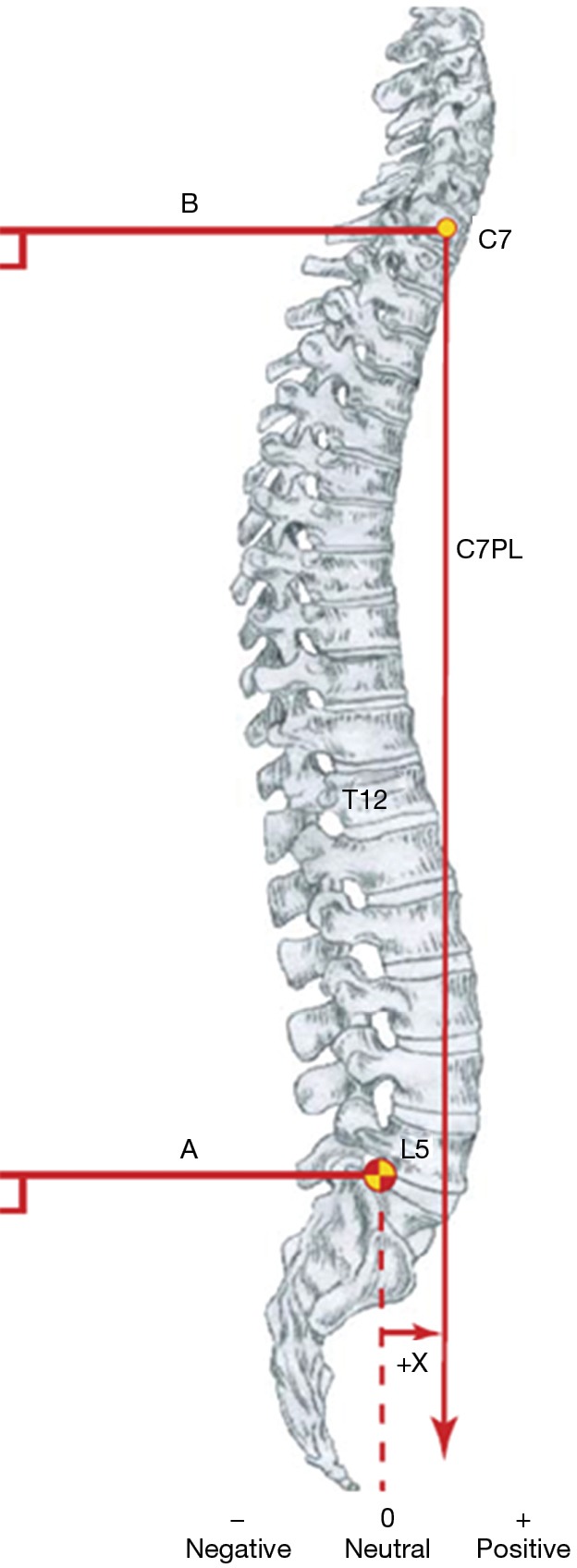
Figure 5.

Sagittal balance. The center of C7 vertebra is marked, and a plumb line has dropped perpendicular to the vertical edge of the radiograph. The posterosuperior corner of S1 is also marked, and a line perpendicular to the vertical edge of the radiograph is constructed. The horizontal difference measured in millimeters between these two lines gives the sagittal balance. No difference between the lines equals sagittal balance. Migration of the line to the front is marked as a positive value, while the movement to the back is negative. Image courtesy of Scoliosis Research Society, from (28).
