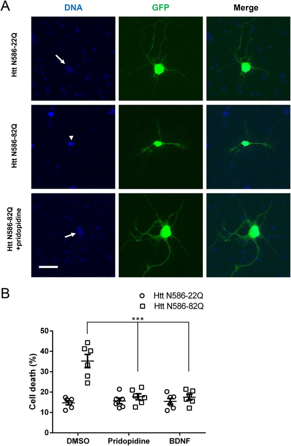
Fig. 1. Pridopidine protects striatal neurons from mutant Huntingtin induced toxicity.
(A) Representative images of GFP transfected striatal neurons. Cells were transfected as stated, treated for 48 h, fixed using 4% paraformaldehyde in PBS, and stained with Hoechst. Healthy non-condensed nuclei (arrow) and apoptotic condensed nuclei (arrowhead) are indicated. Scale bar: 25 μm.
(B) Quantification of nuclear condensation assay in striatal neurons. CD1 primary striatal neurons transfected at DIV5 were treated with 1 μM pridopidine or 20 ng/mL BDNF in the culture media for 48 h before nuclei were stained with Hoechst. Quantification of nuclear staining intensity in transfected cells was performed using Volocity. Results presented as individual values plus means ± S.E. of the percentage of dead cells. *** p < .001 vs DMSO, ANOVA with Bonferroni post-hoc test. (n = 6 independent neuronal preparations).

