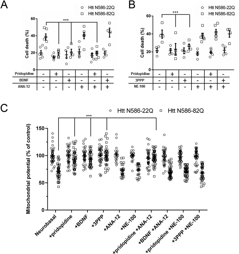
Fig. 4. A sigma-1 receptor antagonist blocks pridopidine-dependent protection of cortical neurons.
Quantification of cell death in neurons treated with pridopidine in combination with a TrkB (ANA-12 1 μM (A)) or sigma-1 receptor (NE-100 1 μM (B)) antagonist. Transfected CD1 primary cortical neurons were treated with a given compound combination in the culture media for 48 h before nuclei were stained with Hoechst. Quantification of nuclear staining intensity in transfected cells was performed using Volocity. Results presented as individual values plus means ± S.E. of the percentage of dead cells. *** p < .001 vs Htt-82Q untreated cells. ANOVA with Bonferroni post-hoc test. (n = 6 independent neuronal preparations).
(C) Quantification of mitochondrial potential using TMRM staining, Average intensity of staining in the soma of transfected cells was measured after 24 h of the indicated treatments. * p < .05 vs Htt N86-82Q by ANOVA with Bonferroni post-hoc test. (n = 4 independent neuronal preparations for a total of 40 independent cells).

