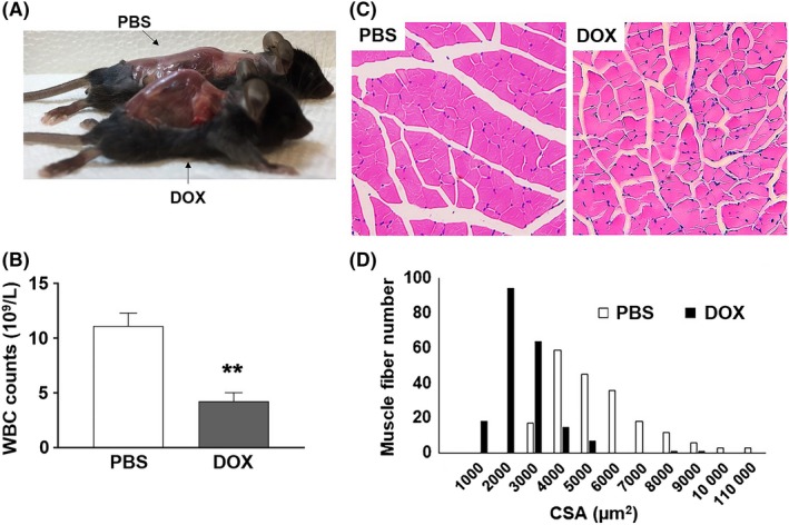
Figure 2.

A, A photograph of skinned PBS‐ or DOX‐treated mouse that exhibits pronounced kyphosis. B, White blood cell (WBC) counts in 16‐18‐wk‐old PBS‐ or DOX‐treated female C57BL/6 mice (n = 4, **P < .01). C, H&E‐stained sections of hind leg skeletal muscle from 18‐wk‐old PBS‐ and DOX‐treated female littermates. D, Frequency distribution of skeletal muscle fiber cross‐sectional area (CSA) between PBS‐ and DOX‐treated mice
