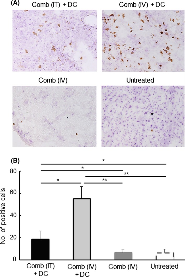
Figure 5.

Effect of IFNγ and CD40L (Comb) gene‐transfection therapy on the infiltration of CD8+ cells into LM8 tumors. Tumor tissue was collected from mice with the indicated treatments at 7 d after the second treatment. Immunohistochemistry was performed to detect CD8+ cells present in the tumor tissue. (A) Typical photos of tumors collected from the indicated treatment groups are shown. Expression of CD8 is shown as brown color products. (B) Counts of CD8+ cells in 1000 cells in tumors are shown. Results are expressed as mean ± SE. The experiment was performed using three mice in each treatment group. All mice were humanely euthanized according to the criteria in Section 2. *P < .05, **P < .01 by the Tukey‐Kramer test
