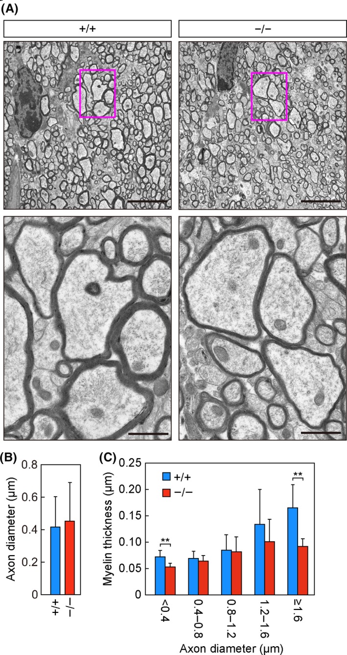
Figure 3.

Thinning of the myelin sheath in Elovl1 −/− Tg(IVL‐Elovl1) mice. A–C) Transmission electron microscopy analysis of the corpus callosum in Elovl1 +/+ Tg(IVL‐Elovl1) and Elovl1 −/− Tg(IVL‐Elovl1) mice at 13 months of age. A) Representative images of the sagittal sections of the corpus callosum at low (5 µm, upper) or high (1 µm, lower) magnification. Red rectangles in upper panels indicate the area enlarged in lower panels. B) Average diameters ± SD of myelinated axons: n = 96 [Elovl1 +/+ Tg(IVL‐Elovl1)] and n = 73 [Elovl1 −/− Tg(IVL‐Elovl1)] total axons counted in 13 different fields of view. C) Myelin thickness of axons counted in (B) was measured and presented according to axon diameter. Values represent the means ± SD. Asterisks indicate significant differences based on Student's t test (**, P < .01). +/+, Elovl1 +/+ Tg(IVL‐Elovl1) mice; −/−, Elovl1 −/− Tg(IVL‐Elovl1) mice
