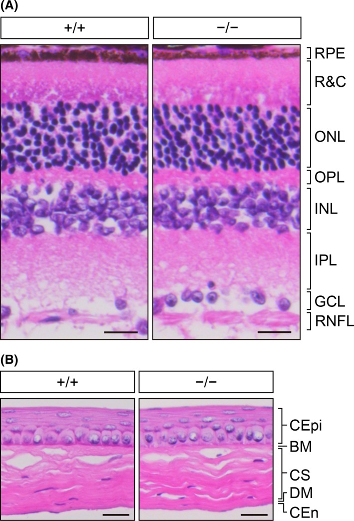
Figure 6.

Normal retinal and corneal histology in Elovl1 −/− Tg(IVL‐Elovl1) mice at 16 weeks of age. Representative hematoxylin and eosin staining images of paraffin sections of the retina (A) and the cornea (B) from 16‐week‐old Elovl1 +/+ Tg(IVL‐Elovl1) and Elovl1 −/− Tg(IVL‐Elovl1) mice. Scale bars, 20 μm. Abbreviations: (A) GCL, ganglion cell layer; INL, inner nuclear layer; IPL, inner plexiform layer; ONL, outer nuclear layer; OPL, outer plexiform layer; R&C, layer of rods and cones; RNFL, retinal nerve fiber layer; RPE, retinal pigment epithelium. (B) BM, Bowman's membrane; CEn, corneal endothelium; CEpi, corneal epithelium; CS, corneal stroma; DM, Descemet's membrane. +/+, Elovl1 +/+ Tg(IVL‐Elovl1) mice; −/−, Elovl1 −/− Tg(IVL‐Elovl1) mice
