Figure 8.

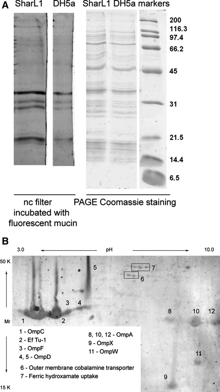
Mucin‐adsorbing membrane proteins from the SharL1 isolate. (A) Mucin‐adsorbing membrane proteins from SharL1 and DH5α. The left panel represents a fluorescent image of a nitrocellulose filter with transferred proteins after separation by PAGE and incubation with a fluorescent‐labelled mucin. The right panel shows proteins separated by 10% SDS/PAGE and Coomassie‐stained. (B) Fluorescent image of a filter with 2D‐PAGE‐separated proteins after incubation with a fluorescent‐labelled mucin. The identified protein spots are indicated. Protein identification details are provided in File S4. Comparison of total bacterial proteome and membrane fraction protein composition is shown in Files S5 and S6
