Figure 1.

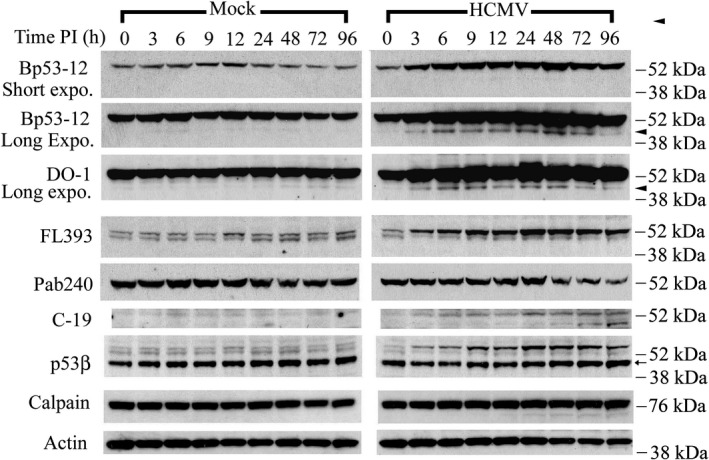
Identification of sub‐53–kDa polypeptides in HCMV‐infected cells, as revealed by a panel of p53 antibodies. Permissive human lung fibroblasts were density arrested and infected with HCMV (5 PFU/cell) or mock infected. Whole‐cell lysates were prepared at the indicated times post infection (PI). Protein aliquots (40 μg/lane) were resolved and membranes probed with the indicated p53 antibodies. Membranes were reprobed with antibody to m‐calpain or actin. These results are representative of at least two independent biological replicates, each one in two technical replicates. Expo: exposure. Arrowhead: p53(ΔCp44). Arrow: p53β. kDa sizes: the relevant molecular weight markers
