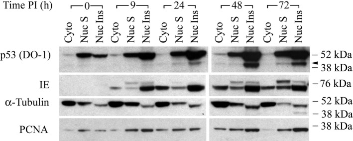
Figure 6.

Subcellular distribution of p53 fragments during the progression of HCMV infection. Density‐arrested LU cells were HCMV‐infected (5 PFU/cell) as in Figure 1. Cells were harvested at the indicated time post infection (PI) and fractionated into a cytosolic fraction (Cyto), Buffer C‐soluble nuclear fraction (Nuc S), and Buffer C‐insoluble nuclear fraction (Nuc Ins). Polypeptides (40 μg/lane) were then analyzed by immunoblot with DO‐1 antibody. Blots were stripped and reprobed with antibody against HCMV IE protein, α‐tubulin, or PCNA. The results are representative of at least two independent biological replicates, each one in two technical replicates. Arrowhead: p53(ΔCp44). kDa sizes: the relevant molecular weight markers
