Figure 1.

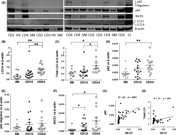
Increased p62, LC3‐II, total LC3, and BICD1 proteins in chronic obstructive pulmonary disease (COPD) patients. Lung tissue from surgical resections were obtained from four healthy volunteers (HV), 10 non‐COPD smoker volunteers (SM), 16 mild CD1/2 (COPD GOLD stage 1 plus GOLD stage 2), and 12 severe CD G3/4 (COPD GOLD stage 3 plus GOLD stage 4) and whole cell extracts were prepared for Western blot. (A) Representative blot. Ct: Control whole cell extract from BEAS‐2B cells. Immunoblotting for p62, LC3‐II, and BICD1 were performed and relative protein amounts were calculated and plotted on a graph against β‐actin: (B) LC3‐II, (C) LC3‐I + LC3‐II (total LC3) (D) p62, (E) p62 oligomers, and (F) BICD1. (G) Correlation between p62 and BICD1 in all patients. (H) Correlation between total LC3 and BICD1 in all patients. Data were analyzed by using Kruskal‐Wallis followed by Mann‐Whitney. P < .05 was considered statistically significant. *P < .05, **P < .01. Whole cell extracts from BEAS‐2B cells were used as control (Ct) in the Western blot membrane
