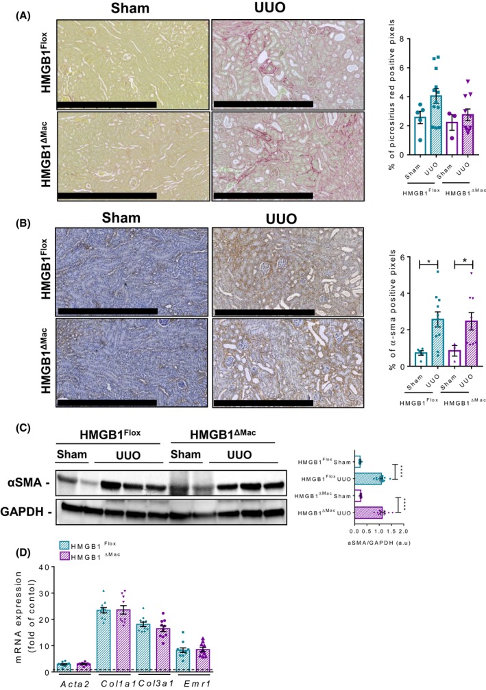
Figure 5.

Macrophage‐specific deletion of high mobility group B1 (HMGB1) does not play a role in UUO‐induced kidney fibrosis. Representative pictures of picrosirius red staining of HMGB1Flox mice and HMGB1ΔMac of kidney section and quantification of positive pixels per kidney section. Scale bar: 500 μm (A). Representative pictures of immunohistochemical staining with an antibody against α‐smooth muscle actin (α‐SMA) and quantification of positive pixels per kidney section. Scale bar: 500 μm (B). Kidney extracts from HMGB1Flox and HMGB1ΔMac mice were analyzed by western blotting directed against α‐SMA (C). Kidney mRNA expression levels of classical fibrosis markers were detected using real‐time RT‐PCR, the dotted line indicates the baseline (D). Statistical analysis was performed with Mann‐Whitney test. Data are expressed as means ± SEM. n = 3 in HMGB1Flox‐sham group; n = 3 in HMGB1ΔMac‐sham group; n = 12 in HMGB1Flox‐UUO group; n = 10 in HMGB1ΔMac‐UUO group. *P < 0.05, **P < 0.01, ***P < 0.001 vs sham/UUO
