
Figure 1.
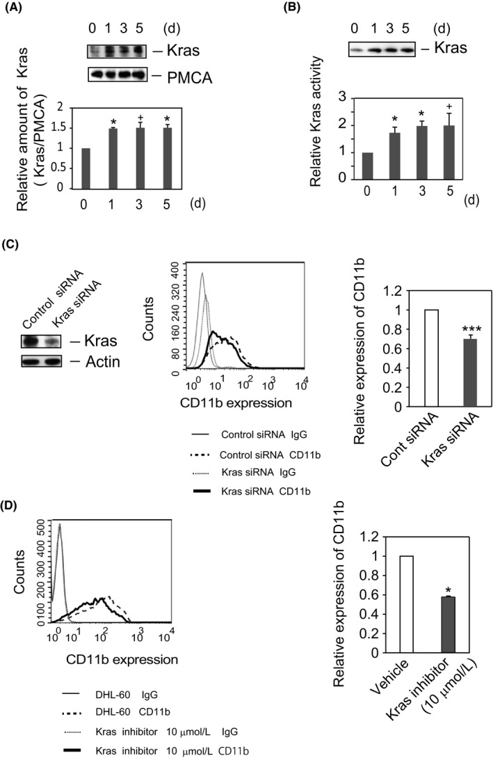
Kras positively regulates the DMSO‐induced differentiation of HL‐60 cells. (A) The amount of Kras is increased in plasma membranes during differentiation of HL‐60 cells. HL‐60 cells were cultured in RPMI medium, with or without 1.3% DMSO, for the indicated times. Plasma membrane fractions were prepared, subjected to SDS‐PAGE, and analyzed by Western blotting with antibody to Kras, followed by incubation with anti‐PMCA antibody, as described in the Materials and Methods. The blots shown are representative of five independent experiments (upper panel). Quantified Kras levels (Kras/PMCA) are displayed as ratio to those in HL‐60 cells (time = 0) and shown as the means ± SE of five independent experiments. *P < 0.05, + P < 0.1. (B) Kras is activated in DHL‐60 cells. Kras activity was assayed using Kras Activation Assay Kits. HL‐60 or DHL‐60 cell lysates were incubated with Raf1 RBD agarose to capture the active form of Kras. The amounts of bound proteins were analyzed by Western blotting with anti‐Kras antibody. A representative blot of four independent experiments is shown (upper panel). Active Kras was detected by immunoblotting with anti‐Kras antibody. The amounts of the active form of Kras were calculated relative to the numbers of cells, and the time course of the ratio of the normalized amount of activated Kras relative to that of resting cells (HL‐60 cells at t = 0) was determined. Quantified results are shown as means ± SE of four independent experiments. *P < 0.05, + P < 0.1. (C) Knockdown of Kras attenuates CD11b expression in DHL‐60 cells. HL‐60 cells were transfected with the indicated siRNA and cultured in RPMI medium containing 1.3% DMSO for 4 days. Cell lysates were analyzed by Western blotting with anti‐Kras or anti‐actin antibody. The blot shown is representative of eight independent experiments (left panel). CD11b expression was analyzed by flow cytometry. Data representative of four independent experiments are shown (middle panel). IgG; normal IgG‐staining cells. The ratio relative to the cells transfected with control siRNA is shown as mean ± SE of four independent experiments (right panel). ***P < 0.005. (D) Kras inhibitor attenuates the expression of CD11b in DHL‐60 cells. Cells were cultured in RPMI medium containing DMSO (1.3%) with or without Kras inhibitor (10 μmol/L) for 5 days. CD11b expression was analyzed by flow cytometry. Data representative of four independent experiments are shown (left panel). The ratio of the geometric mean of CD11b expression on the cells treated with Kras inhibitor to that on cells treated with vehicle is shown as the mean ± SE of four independent experiments (right panel). *P < 0.05