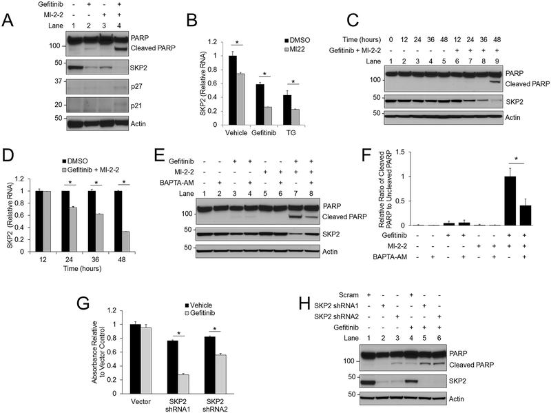
Figure 5: Gefitinib and menin inhibition synergistically decrease SKP2 expression in a calcium-dependent manner.
A) HT-29 cells treated for 48 hours followed by analysis of protein levels by western blotting. 10 μM gefitinib, 1 μM MI-2–2. B) After 48 hours of treatment in HT-29 cells SKP2 mRNA was assessed by RT-PCR and plotted relative to actin. 1 μM MI-2–2,10 μM gefitinib, 5 nM TG. C-D) A time course was performed in HT-29 cells treated with either vehicle or 10 μM gefitinib/1 μM MI-2–2, with analysis of protein levels by western blotting (C) and SKP2 mRNA assessment by RT-PCR, plotted relative to actin, and normalized to DMSO for each time point (D). E) HT-29 cells treated for 48 hours with protein analysis by western blot. 10 μM gefitinib, 1 μM MI-2–2, 5 μM BAPTA-AM. F) Quantitation and normalization of cleaved and uncleaved PARP levels on western blot from Figure 5E. G-H) HT-29 cells transduced with either scrambled or SKP2 shRNAs, then treated with 10 μM gefitinib. After 96 hours, cell growth was assessed by the MTS assay (G) and after 48 hours protein levels were assessed by western blot (H). * p < 0.05.

