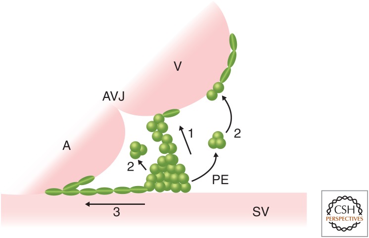
Figure 2.
An overview of proepicardial organ (PE) translocation to the myocardium. Schematic of PE translocation to the myocardium showing three models across species. (1) Direct contact: In the chick, Xenopus, axolotl, zebrafish, and mouse, the PE produces protrusions to form a tissue bridge between the PE and the dorsal wall of the ventricle (or the PE contacts the ventricle directly) to allow the PE cells to migrate on to the myocardium. (2) Floating cysts: In zebrafish and mice, PE cell clusters detach from the proepicardial surface to form cysts, which float across the pericardial cavity to attach to the ventricular wall. (3) Migration: In mice, PE cells migrate from the SV toward the heart along the surface of the inflow tract. A, Atrium; V, ventricle; AVJ, atrioventricular junction; SV, sinus venosus.

