-
A, B
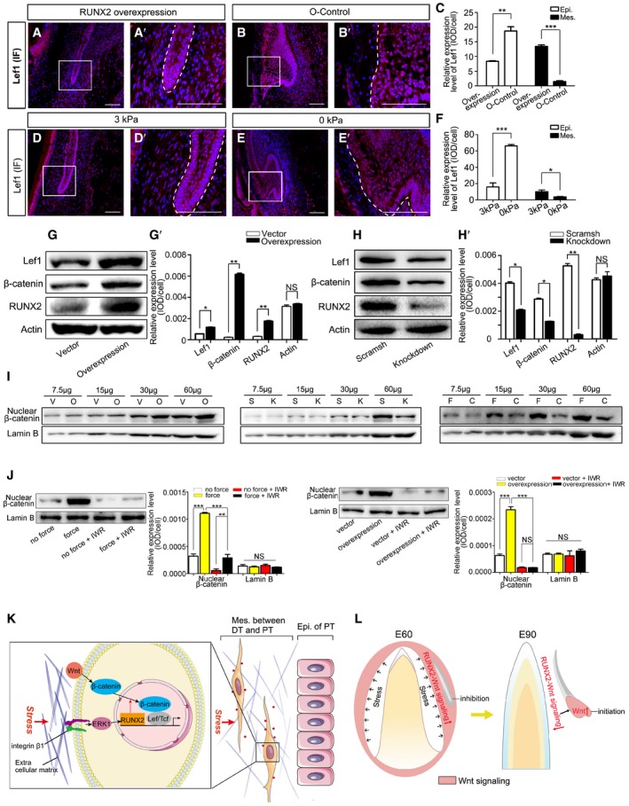
IF of Lef1 in mandible slices at embryonic day 60 (E60) infected with RUNX2 overexpression lentiviral vector (overexpression) or overexpression control lentiviral vector (O‐control). (A’–B’) are magnifications of boxed regions in the corresponding figure panels. Dashed lines mark the epithelium of PC.
-
C
Relative IF expression levels of Lef1 in epithelium (Epi.) and mesenchyme (Mes.) of the overexpression and O‐control groups.
-
D, E
IF of Lef1 in E60 mandible slices subjected to 3 or 0 kPa stress for 2 days. (D’, E’) are magnifications of boxed regions in the corresponding figure panels. Dashed lines mark the epithelium of PC.
-
F
Relative IF expression levels of Lef1 in the epithelium and mesenchyme after applying 3 and 0 kPa pressure.
-
G
Western blots of Lef1, non‐phospho‐β‐catenin, and RUNX2 after DFCs were infected with control lentiviral vector or RUNX2 overexpression lentiviral vector. (G’) Relative expression levels between control vector and RUNX2 overexpression groups.
-
H
Western blots of Lef1, non‐phospho‐β‐catenin, and RUNX2 after DFCs were infected with scrambled shRNA (scramsh) or RUNX2 knockdown shRNA (knockdown) lentiviral vectors. (H’) Relative expression levels between scramsh and knockdown groups.
-
I
Western blots of nuclear non‐phospho‐β‐catenin and Lamin B in DFCs of RUNX2 overexpression (O), control vector (V), RUNX2 knockdown (K), scramsh (S), compressed force (F; 1.0 g/cm2), and control (C; 0 g/cm2) groups. Sample was loaded in a gradient (7.5, 15, 30, and 60 μg) showing a linear relationship for each group.
-
J
Western blots of nuclear non‐phospho‐β‐catenin and Lamin B in DFCs treated with IWR‐1‐endo. Relative expression levels were compared between force and no force groups and between overexpression and control groups with or without IWR‐1‐endo treatment.
-
K
Diagram illustrating the biomechanical stress regulation of Wnt/β‐catenin signaling in the mesenchyme between the deciduous (DT) and permanent tooth (PT) via the integrin β1‐ERK1‐RUNX2‐Wnt/β‐catenin pathway.
-
L
Diagram illustrating the biomechanical stress‐associated downregulation of RUNX2‐Wnt/β‐catenin pathway in the mesenchyme, inducing upregulation of Wnt signaling in the epithelium, which triggers PT development.
Data information: Data represent the means ± SEM. Scale bars = 100 μm.
= 3 for all experiments. Unpaired
‐tests for (C, F, G’, and H’); one‐way ANOVA (Newman–Keuls test for post hoc comparisons between two groups) for (J), *
0.001; NS. not significant.

