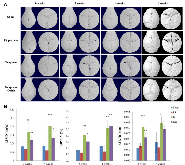
Figure 4.
Micro-CT imaging analysis of murine calvarial model treated with different particles. (A) Reconstructed image of whole skull and VOI with the midline suture of the skull. The VOI is defined with a diameter of 5 mm. (B) Bone resorption parameter quantified by micro-CT in calvarial tissues (mean ± SD, *p < 0.05, **p < 0.01, ***p < 0.001).

