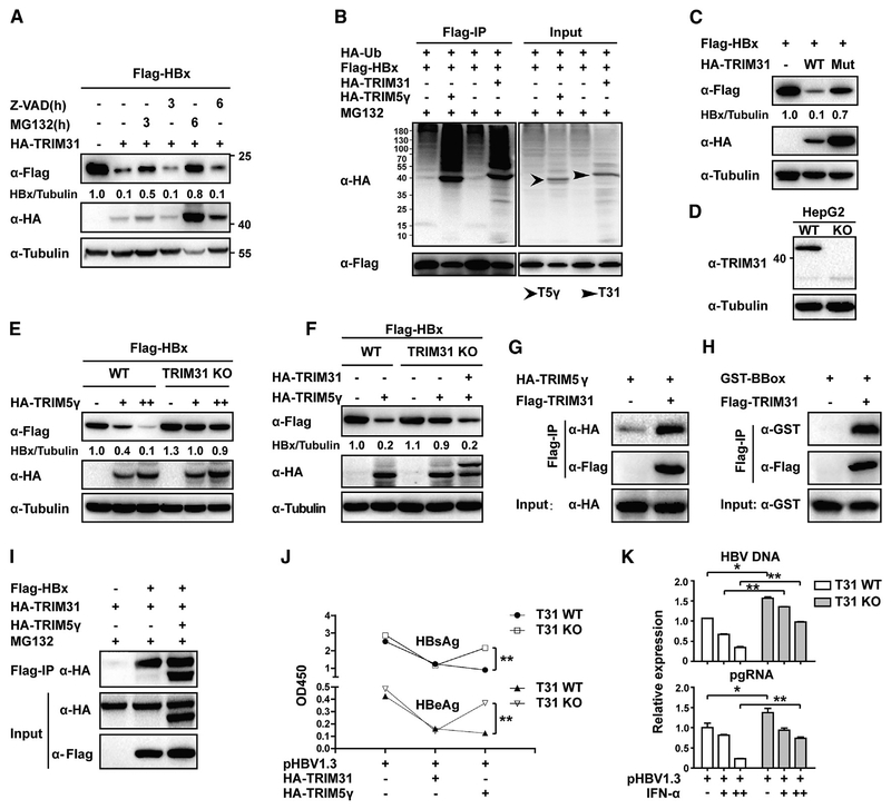
Figure 7. TRIM5γ Recruits TRIM31 to Degradate HBx.
(A) HepG2 cells were co-transfected with FLAG-HBx and HA-TRIM31 or HA empty vector plasmids; 36 h later, cells were left untreated or were treated with MG132 (10 μM) or Z-VAD (10 μM) as indicated. Cells were harvested after treatment, and whole-cell lysates were immunoblotted with the indicated antibodies. (B) HepG2 cells were transfected with FLAG-HBx, HA-Ub, HA-TRIM5γ, or TRIM31 expression plasmids as indicated; 24 h later, cells were treated with MG132 for 6 h and were collected and subjected to coIP analysis. (C) HepG2 cells were transfected with FLAG-HBx or co-transfected with TRIM31 plasmids (WT or mutant); 36 h later, cells were harvested and analyzed as in (A). (D) HepG2 WT or TRIM31 KO cell lysates were immunoblotted with TRIM31 or Tubulin antibody. (E) HepG2 WT orTRIM31KO cells were transfected withFLAG-HBxwithor without the TRIM5γ expression vector; 28h later, cells were collected and analyzed asin(A). (F) WT HepG2 cells or TRIM31 KO cells were transfected as indicated; 28 h later, cells were collected and analyzed as in (A). (G and H) Expression vectors for HA-TRIM5γ (G) or GST-BBox (H) were co-transfected into 293T cells with or without FLAG-TRIM31; 36 h later, cells were subjected to coIP using FLAG-TRIM31. Immunoblot analyses were carried out using anti-FLAG, anti-HA, or anti-GST antibody. (I) 293T cells were transfected with FLAG-HBx, HA-TRIM5γ, or HA-TRIM31 expression plasmids as indicated; 24 h later, cells were treated with MG132 (10 μM) for 8 h and subjected to coIP analysis. Immunoblotting was carried oout using an anti-FLAG or anti-HA antibodies. Data are representative of at least three independent experiments. (J) HepG2 WT or TRIM31 KO cells were transfected with pHBV1.3 plasmids or together with TRIM5γ or TRIM31 expression plasimds as indicated; 72 h later, the supernatant was tested for HBeAg and HBsAg content using ELISA. Mean ± SD values from three independent experiments are shown. **p < 0.01. (K) HepG2 WT or TRIM31 KO cells were transfected with pHBV1.3 plasmids; 24 h later, cells were treated with IFN-α (10ng/ml) or untreated. After another 48 h, qPCR was performed to evaluate the HBV pgRNA and DNA levels. Mean ± SD values from three independent experiments are shown. *p < 0.05, **p < 0.01.

