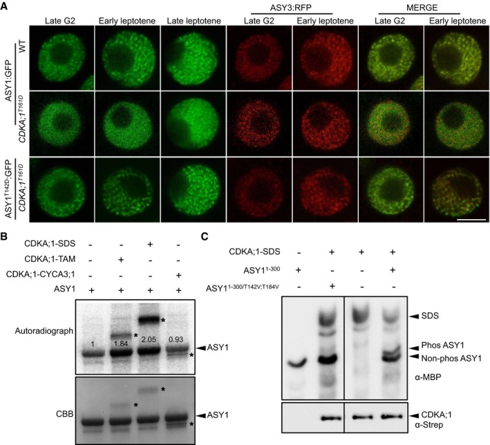
Figure 2. ASY1 is a phosphorylation target of CDKA;1.

- ASY1:GFP and ASY1T142D:GFP localization in late G2 and leptotene of male meiocytes of the wild‐type and CDKA;1 T161D mutants. ASY3:RFP, highlighting chromosomes, was used as a marker for the staging of meiosis. Scale bar: 5 μm.
- Kinase assays of CDKA;1‐SDS, CDKA;1‐TAM, and CDKA;1‐CYCA3;1 complexes using ASY1 purified from baculovirus‐infected insect cells as a substrate. The upper panel shows the autoradiograph. The control reaction without CDKA;1–cyclin complex indicates a background activity co‐purified from insect cells. The lower panel indicates protein loading by Coomassie Brilliant Blue (CBB) staining. Arrowheads indicate ASY1 proteins, and asterisks depict the relevant cyclin used which also gets phosphorylated in the assay. Numbers indicate the relative intensities of ASY1 bands.
- The upper panel shows a phos‐tag gel analysis of ASY11–300 and ASY11–300/T142V;T184V with and without CDKA;1‐SDS kinase complexes using an anti‐MBP antibody. The lower panel denotes loading of CDKA;1 using an anti‐Strep antibody. Arrowheads represent the proteins as indicated.
Source data are available online for this figure.
