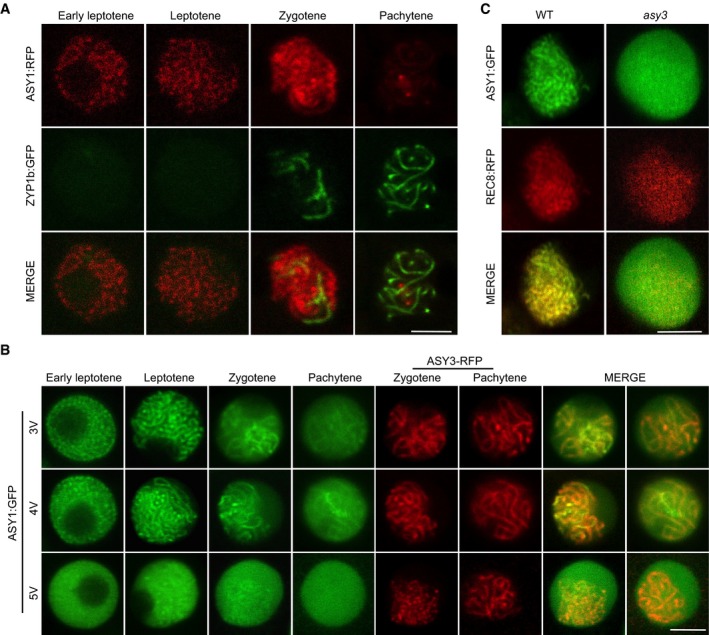
Figure EV2. Localization of ASY1 variants in the wild‐type and asy3 mutants.

- Co‐localization analysis of ASY1‐RFP with ZYP1b‐GFP at different meiotic stages in male meiocytes of the wild type. Scale bars: 5 μm.
- Localization of ASY13V:GFP (T365V S382V T535V), ASY14V:GFP (T184V T365V S382V T535V), and ASY15V:GFP (T142V T184V T365V S382V T535V) together with ASY3:RFP (for staging of zygotene and pachytene) at different meiotic stages in male meiocytes of asy1 mutants. Scale bars: 5 μm.
- Localization of ASY1:GFP in the male meiocytes of the wild‐type and asy3 mutants at leptotene. REC8‐RFP was used for staging and to highlight chromosomes. Scale bars: 5 μm.
