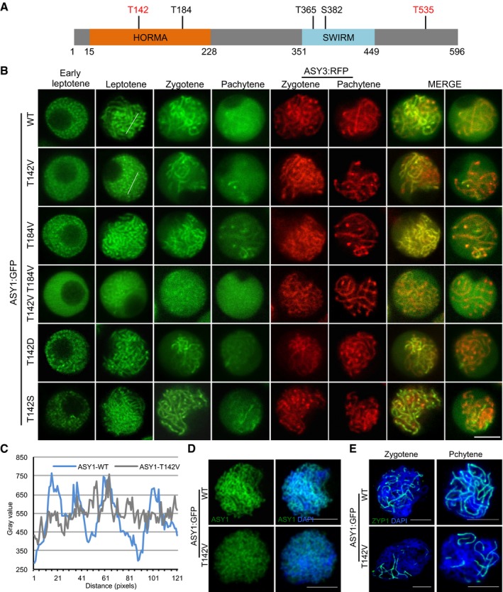
Figure 3. Phosphorylation of ASY1 is essential for its chromosomal localization.

-
ASchematic representation of ASY1 with the five predicted consensus Cdk phosphorylation sites. The sites found to be phosphorylated in vitro by CDKA;1‐SDS complexes are highlighted in red (Appendix Fig S4A).
-
BLocalization patterns of different ASY1:GFP variants together with ASY3:RFP (for staging of zygotene and pachytene) in a asy1 mutant background during prophase I. Scale bar: 5 μm.
-
CSignal distribution profiles of ASY1:GFP and ASY1T142V:GFP at leptotene as shown in (B). The regions used for analysis are highlighted by white lines in respective panels in (B). The many small peaks with low amplitude in ASY1 T142V :GFP indicate diffused localization as opposed to the clear peaks seen in the wild type.
-
D, EImmunolocalization of ASY1 (D) and ZYP1 (E) in ASY1:GFP (asy1) and ASY1 T142V :GFP (asy1) plants using anti‐GFP and anti‐ZYP1 antibodies, respectively. DNA was stained with DAPI (blue). Scale bars: 5 μm.
