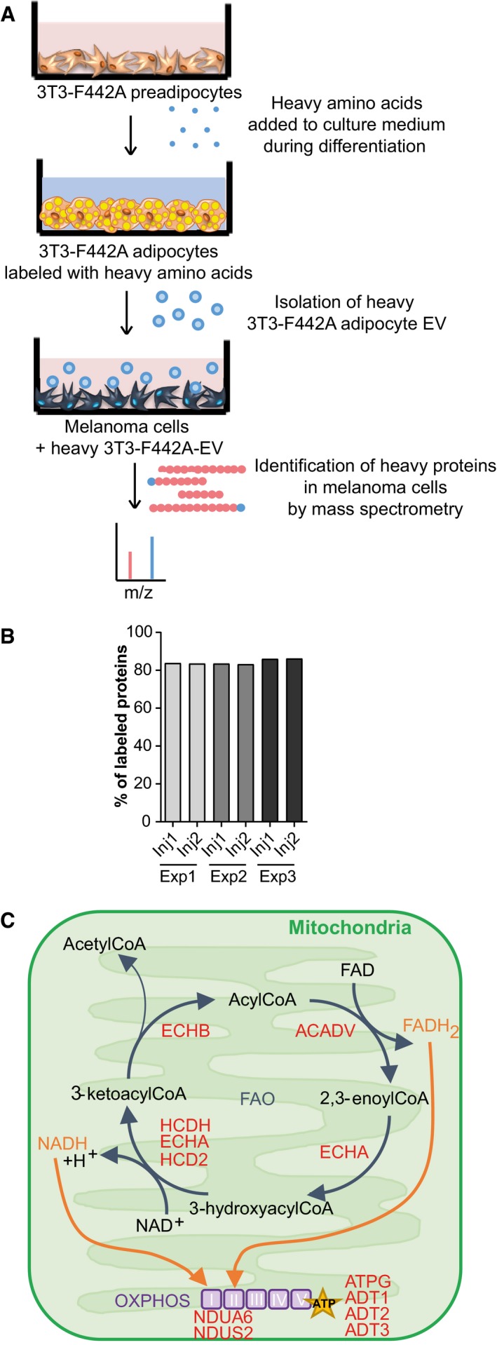
Figure 1. Adipocyte EV transfer proteins involved in FA metabolism to melanoma cells.

- Workflow of the SILAC approach. 3T3‐F442A cells were seeded and differentiated in the presence of heavy amino acids. After 14 days of differentiation, the EV secreted by the mature labeled adipocytes were isolated and analyzed by mass spectrometry to evaluate the presence of heavy amino acid‐containing proteins. These EV were also added to SKMEL28 cells for 12 h, and then, LC‐MS/MS analysis was performed to identify heavy amino acid‐containing proteins that had been transferred from adipocytes to melanoma cells via EV.
- Three independent samples (Exp 1–3) of EV secreted by labeled 3T3‐F442A cells were analyzed by mass spectrometry (in duplicate injections, Inj1/2). The percentage of proteins bearing at least one peptide containing a heavy amino acid is indicated.
- Proteins involved in FAO and oxidative phosphorylation (OXPHOS) that are transferred from adipocytes to melanoma cells via EV are shown in red.
