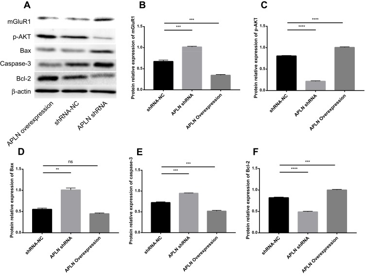
Figure 3.
Influence of APLN on the expression of apoptosis-associated proteins, mGluR1, and p-AKT levels. Hippocampus neurons of epileptic models were transfected with pBI-CMV3-APLN overexpression, short hairpin RNA negative control (shRNA-NC), or interference APLN shRNA plasmids. Forty-eight hours post-transfection, protein expression was evaluated by Western blotting. β-actin was used as a loading control. (A) Western blotting was used to examine the protein levels of mGluR1, p-AKT, Bax, caspase-3, and Bcl-2. Statistical results of mGluR1 expression (B), p-AKT levels (C), Bax expression (D), caspase-3 expression (E), and Bcl-2 expression (F) in different groups. ns, non-significant; **P < 0.01, ***P < 0.001, ****P < 0.0001.

