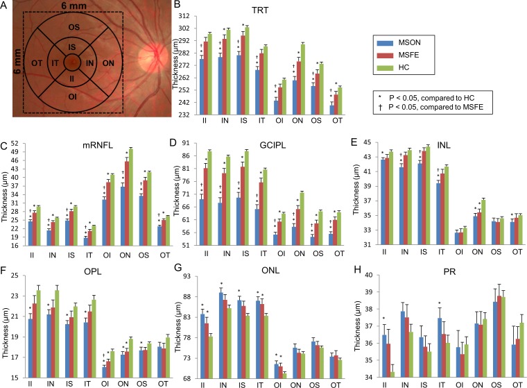
Figure 7.
Sectoral thickness differences of the intraretinal layers in patients with MS compared with controls. (A) According to the ETDRS definition, the quadrantal division was performed using 45° and 135° medians. Three concentric rings with diameters of 1, 3, and 6 mm were used to divide the map into nine zones. The central 1-mm zone of the fovea was removed. Significant thickness reductions were found in the TRT, mRNFL, and GCIPL in MSON compared with those of MSFE and HC in sectoral partitions using GEE models (P < 0.05). (B–H) GCIPL thickness in each sector of TRT and six retina layers. II, inner inferior; IN, inner nasal; IT, inner temporal; OI, outer inferior; ON, outer nasal; OT, outer temporal; OS, outer superior. Bars = SEs.

