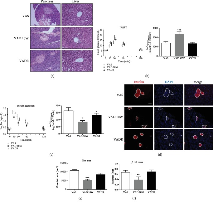
Figure 2.
VAD leads to irregular islet morphology, loss of β cell mass, and impaired glycemic responses. (a) H & E staining of pancreatic islet and liver sections of mice from VAS, VAD, and VADR groups. Magnification, 40x; scale bars, 50 μm. (b, c) Blood glucose and insulin levels using the IPGTT test were analyzed of mice described for (a). The results of the IPGTT test were analyzed by AUC (i.e., AUCIPGTT-glucose or AUCIPGTT-insulin). (d) Representative immunofluorescence images of pancreatic islet sections of mice described for (a) stained with antibodies against insulin. Magnification: 20x; scale bars, 100 μm. (e) Mean islet area (μm2) of pancreas of mice described for (a). (f) β cell mass of mice described for (a). Error bars represent S.E. ∗p < 0.05, ∗∗p < 0.01, and ∗∗∗p < 0.001.

