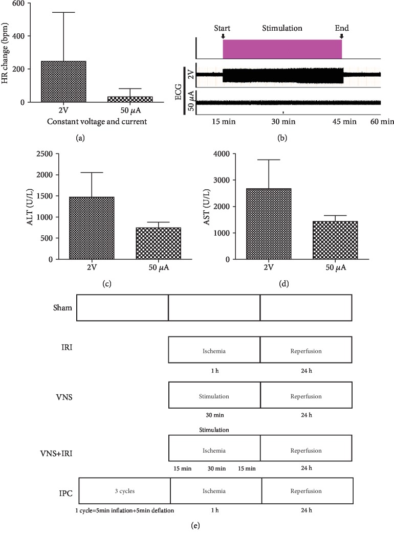
Figure 1.
Establishment and optimization of vagus nerve stimulation. Heart rate was recorded as rats underwent vagus nerve stimulation at given parameters (1 ms, 10 Hz), but with different stimulus intensities (2 V, 50 μA). Changes in the heart rate (a) of vagal stimulation compared with ischemia without stimulation. 50 μA of current reduced heart rates more reliably. (b) Electrocardiograph during left vagal stimulation. (c, d) Rats underwent vagal stimulation or sham during the ischemic period, and blood was collected and tested at the end of 24 h of reperfusion. (e) Experimental protocol and established five experimental groups. ∗∗P < 0.01.

