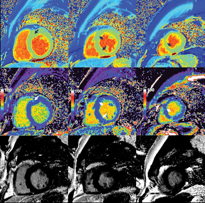
Figure 4:
Native myocardial T1 images (top row), quantitative extracellular volume (ECV) fraction images (middle row), and late gadolinium enhancement (LGE) images (bottom row) in a 62-year-old man with interventricular septal hypertrophic cardiomyopathy. Patchy intramyocardial areas of elevated T1 and ECV were mainly in interventricular septum (arrows). These findings suggest increase of extracellular matrix in involved myocardium. No obvious LGE was observed on conventional LGE images in bottom row.

