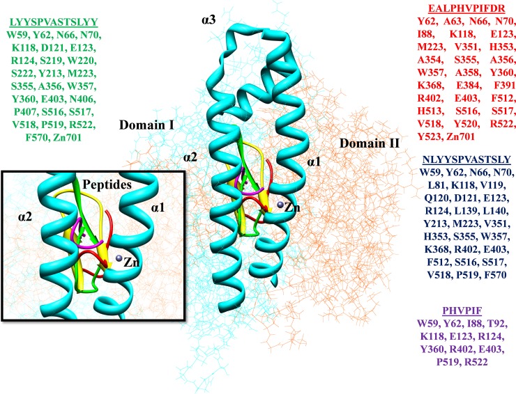
Fig 5. Interacting residues of the ACE-peptides are represented in different colors.
The crystal structure of human ACE (PDB ID: 1O8A) protein is divided into two domains as Domain I (N-terminal) (37–291 amino acids) represented in cyan color while Domain II as C-terminal domain is presented in orange color (292–625 amino acids). The N-terminal lid appeared as the α1, α2, and α3 exhibiting the active site of protein along with the Zn binding site. The scrutinized peptides showed the interactions at binding sites and represented in different colors along with interacting residues.

