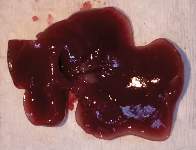
Figure 2a:

Intrahepatic metastatic tumor burden after radiofrequency ablation (RFA). Whole excised livers are presented as viewed from the ventral surface. At 14 days, the number of CT26 tumors in livers of (a) mice undergoing a sham procedure was significantly smaller than that in (b) mice that underwent RFA. Likewise, at 21 days, tumor number for (c) the sham group implanted with MC38 was overwhelmingly exceeded by that of (d) the RFA group. An ellipsoid white area (arrow in d) represents the visualized zone of residual radiofrequency-induced coagulation.
