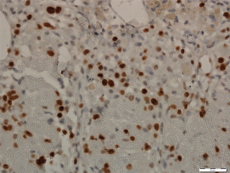
Figure 3d:

Photomicrographs show tumor proliferation induced by radiofrequency ablation (RFA) in mice. Brown dots = Ki-67–positive staining in the cellular nucleus (original magnification, ×40). Mice undergoing RFA developed more highly proliferative tumor cells than their littermates in the sham group. (a) Ki-67 in the sham group of Balb/C mice with CT26. (b) Ki-67 in the RFA group of CT26. (c) Ki-67 in the sham group of C57BL6 mice with MC38. (d) Ki-67 in the RFA group of C57BL6 mice with MC38.
