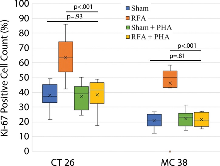
Figure 5c:

Graphs show that tumor load, proliferation, and microvascularization in mice undergoing radiofrequency ablation (RFA) are reduced after the administration of inhibitors of c-Met (ie, PHA) and signal transducer and activator of transcription 3 (S3I). (a, b) The overall tumor load of intrahepatic metastases after RFA declined to baseline with the addition of adjuvant PHA or S3I, compared with that in the group undergoing RFA alone (P < .05), with an overall number equivalent to that in the sham group (P > .05). (c, d) More proliferative tumor cells developed in mice undergoing RFA than in their littermates in the sham group (P < .001 for both CT26 and MC38), with proliferation reduced when RFA was combined with adjuvant PHA or S3I to an equivalent baseline level of the sham group (P > .05 for both tumor models). (e, f) Higher microvascular density was also noted within the tumor in the RFA group (P < .001 compared with sham for both tumor models), whereas lower microvascular density was observed when adjuvant PHA or S3I was administered after RFA and versus RFA alone (P < .001 for both cell lines), with no statistical difference from the sham procedure alone (P > .05 for both cell lines).
