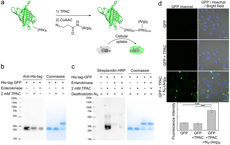
Figure 5.
Functionalization of His-tag on GFP to enable protein delivery. (a) Scheme of bioconjugation of polyarginine onto His-tag of a fluorescent protein using TPAC. (b) Streptavidin-HRP blot and coomassie stain showing that removal of the His-tag significantly reduces the labeling by TPAC. His-tagged GFP and native GFP prepared by enterokinase cleavage of His-tag were treated with TPAC, followed by CuAAC with desthiobiotin-N3 and gel-analysis. Cleavage of His-tag leads to less coomassie staining but with similar migration on SDS-PAGE (Figure S14a). (c) Western blot and coomassie stain showing that labeling of TPAC with His-tagged GFP significantly reduces its detection by His-tag antibody. (d) Transduction of functionalized GFP into live HeLa cells. Cells were incubated with GFP (0.1 μg/μL), stained with Hoechst 33 342 and imaged; quantification is shown on the bottom (n = 3, average ± s.d. ***P ≤ 0.001; two-tailed Student’s t test.). Scale-bars: 20 μm.

