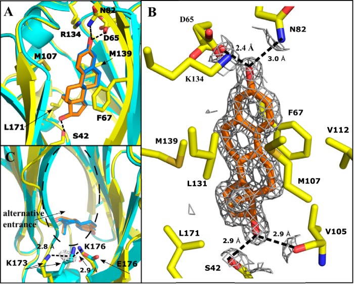
Figure 2.
Crystal structure of estradiol (orange) bound to the LG4 domain of E176K SHBG (yellow) modeled at 1.73 Å resolution. Hydrogen bonds are indicated as dotted lines. Structure of WT SHBG-bound estradiol (PDB: 1LHU) (cyan, with estradiol highlighted blue) is overlaid for comparison. A, overlay of E176K SHBG and WT SHBG with estradiol bound, highlighting the similarity of the two structures and steroid positioning. One cycle alignment RMSD = 1.25 Å across all Cα atoms. All key residue side chains are in the same position for both structures. B, estradiol positioned in the binding pocket of E176K SHBG with residues involved in the binding interaction shown. 2Fo − Fc map is shown around the ligand at 1.5 σ, carved at 3.0 Å. C, proposed alternative entrance to the binding pocket is shown, with the E176K substitution and resulting hydrogen bond interaction with Lys-173 through a water molecule (red) shown.

