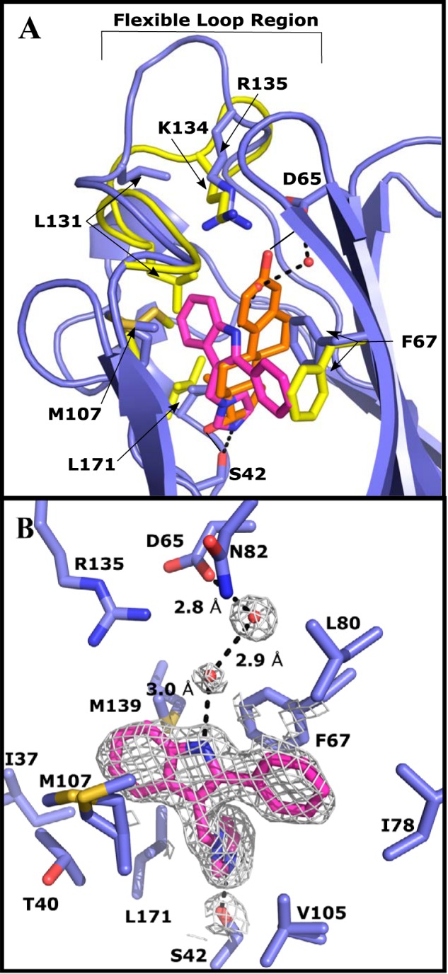
Figure 4.

Crystal structure of IPI (purple) bound to the LG4 domain of E176K SHBG (blue) modeled at 1.71 Å resolution. An overlay of the differences between the crystal structure of E176K estradiol (yellow, estradiol is orange) and the IPI-bound E176K SHBG highlights the key differences around the binding pocket. One cycle alignment RMSD = 1.35 Å across all Cα atoms. Water molecules involved in the binding are shown as red spheres and hydrogen bonds are indicated as dotted lines. A, IPI-SHBG (blue) is aligned with estradiol-SHBG (yellow) with the differences in the flexible loop region and key positions of key residues indicated. IPI and estradiol are compared in the binding site highlighting the differences in the volume occupied by the nonsteroidal ligand. B, IPI binding interactions and the residues forming the hydrophobic pocket are shown. 2Fo − Fc electron density (1.5 σ) is shown around DVT and the water molecules in the binding pocket.
