. 2019 Dec 31;8:e50796. doi: 10.7554/eLife.50796

Figure 6. Cdc7 phosphorylates T916 and S945.

(A) Two hundred ng (34 pmole) each of wild type CKBD peptide (wild-type) or mutant peptides (CKBD-A, Others-A and Others-E) was incubated in the kinase reaction with indicated amounts of purified Cdc7-ASK and/or CK1γ1 in the presence of [γ-32P]-ATP for 60 min at 30°C. Samples were analyzed on 18% SDS-PAGE. After silver staining, the gel was autoradiographed. The position of the substrate peptides in the gel is indicated by an arrow. The amino acid sequences of each peptide are shown below the panel. The serine and threonine in CKBD are in green, and the mutated residues are shown in red. (B) Quantification of the phosphorylation level of the peptide in (A). The averages of three independent kinase assays are shown with error bars.

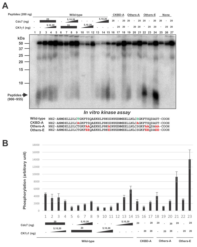
Figure 6—source data 1. Quantification for graph (three independent FACS experiments) in Figure 6B.

Figure 6.

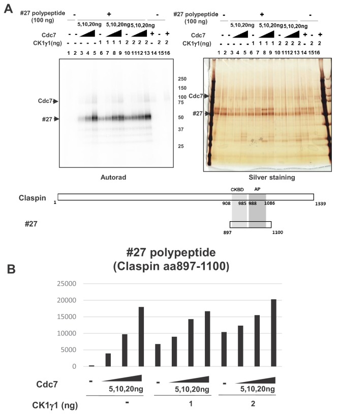
Figure 6—figure supplement 1. Cdc7 and CK1γ1 phosphorylate Claspin independently.

Figure 6—figure supplement 1.

(A) Purified Claspin polypeptide #27 (aa897-1100 containing CKBD and AP) was incubated in the kinase reaction with indicated amounts of purified Cdc7-ASK and/or CK1γ1 in the presence of [γ-32P]-ATP. Samples were analyzed on 4–20% gradient SDS-PAGE, followed by silver staining (right) and autoradiogram (left). The positions of the proteins are indicated by arrows. Bottom drawing shows schematic diagram of the polypeptide #27. (B) Quantification of the phosphorylation level of the #27 polypeptide in A.
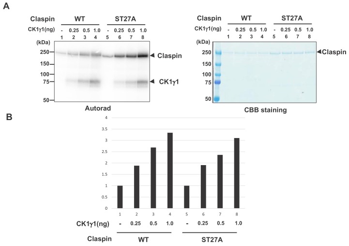
Figure 6—figure supplement 2. CK1γ1 phosphorylates Claspin in a manner independent of serine/threonine residues in the CKBD-AP segment.

Figure 6—figure supplement 2.

(A) Purified wild type Claspin or Claspin mutant ST27A (serines and threonines in aa903-1120 substituted by alanine) was incubated in the kinase reaction with indicated amounts of purified CK1γ1 in the presence of [γ-32P]-ATP. Samples were analyzed on 4–20% gradient SDS-PAGE, followed by CBB staining (right) and autoradiogram (left). The positions of the proteins are indicated by arrows. Bottom drawing shows schematic diagram of the polypeptide #27. (B) Quantification of the phosphorylation levels of the Wt or ST27A Claspin in A. The background phosphorylation level in the absence of CK1γ1 was taken as one for both WT and ST27A Claspin proteins.