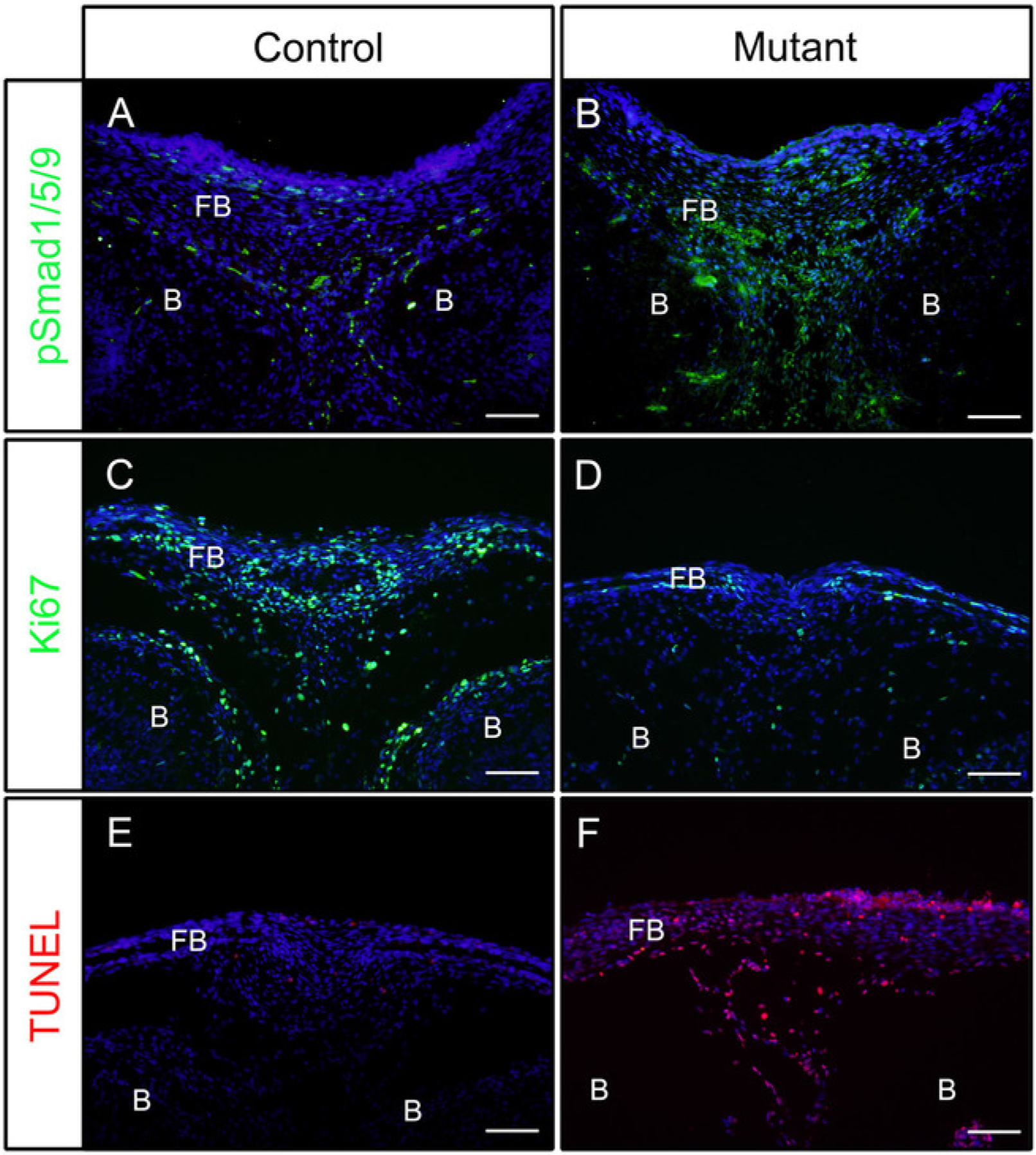
Figure 1: Examples of IF results of pSmad1/5/9, Ki67 or TUNEL in control embryos and mutant embryos with enhanced BMP activity.

Constitutively activated Bmpr1a (caBmpr1a) mice were crossed with P0-Cre mice to increase BMP signaling activity in neural crest cells (NCCs). Heads of control (P0-Cre; caBmpr1a+/+) and mutant (P0-Cre; caBmpr1afx/+) embryos were dissected at E16.5 or E18.5, fixed with 4% PFA for 4h, cryoprotected with 30% sucrose for 1 day, embedded in OCT, and cryosectioned at −18 °C. Sections of the frontal bone (similar level with the eye) were used for immunodetection against pSmad1/5/9, Ki67, or TUNEL staining. (A, B) pSmad1/5/9 (green) staining patterns in the frontal bones of control (A) or mutant (B) embryos at E16.5. (C, D) Ki67 (green) staining patterns in the frontal bones of control (C) or mutant (D) embryos at E18.5. (E, F) TUNEL (red) staining patterns in the frontal bones of control (E) or mutant (F) embryos at E18.5. Nuclei were stained with DAPI (blue). FB = frontal bone, B = brain. Scale bars = 100 μm.
