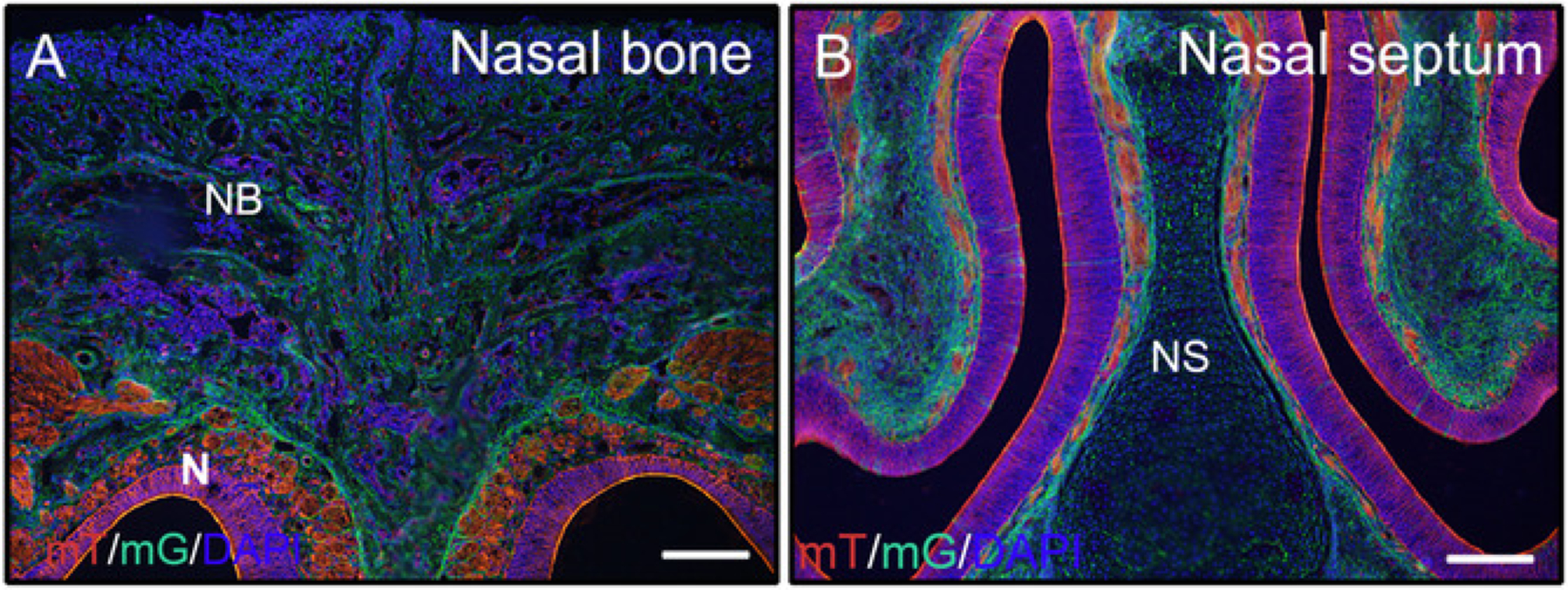
Figure 2: Examples of mTmG reporter signal results of undecalcified tissues in the head.

Heads from 3 week old P0-Cre mice with membrane-tomato and membrane GFP (mTmG) reporter were dissected, fixed with 4% PFA for 4h, cryoprotected with 30% sucrose for 2 days, embedded in 8% gelatin, and cryosectioned at −25 °C. Head sections clearly show GFP (green, Cre recombination positive) and Tomato (red, Cre recombination negative) signal in the nasal bone and nasal tissues (A, B). Nuclei were stained with DAPI (blue). NB = nasal bone, N = nasal tissues, NS = nasal septum. Scale bars = 250 μm.
