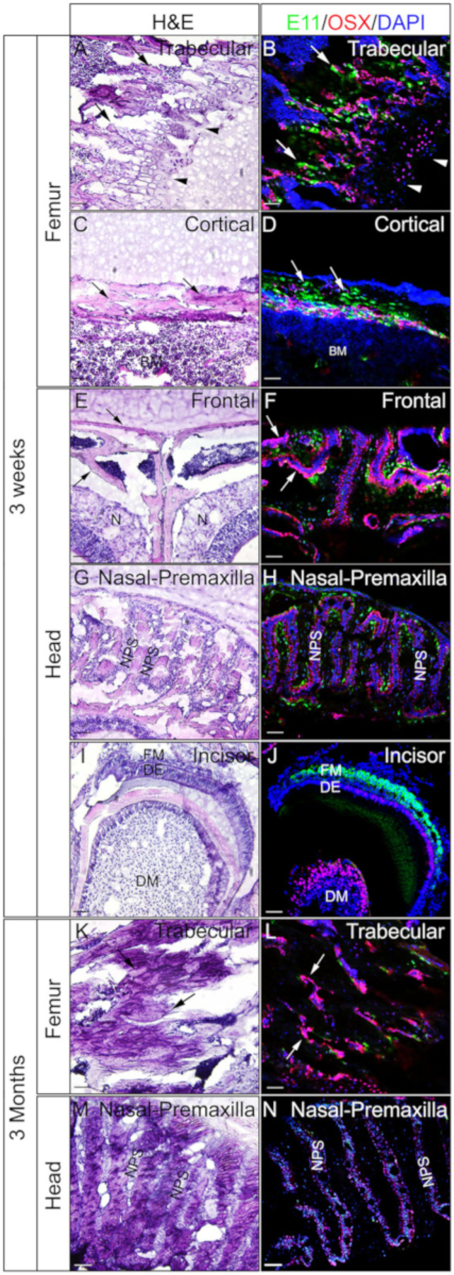
Figure 4: Examples of OSX and E11 double immunostaining results of undecalcified tissues in the head and the femora.

Heads and femora were dissected from 3 week-old or 3 month-old mice, fixed with 4% PFA for 4h, cryoprotected with 30% sucrose for 2 days, embedded in 8% gelatin, and cryosectioned at −25 °C. Sections were used for double immunostaining with antibodies against OSX (Red) and E11/Podoplanin (Green). Nuclei were stained with DAPI (blue) (B, D, F, H, J, L, N). Adjacent sections of those tissues were used for H&E staining (A, C, E, G, I, K, M). Arrows in A, B, K, and L indicate trabecular compartments of the femur; C and D, cortical compartments of the femur; and in E and F, the frontal bones. Arrowheads in A and B indicate growth plate. BM = bone marrow, N = nasal tissues, DM = dental mesenchyme, DE = dental epithelium, FM = follicle mesenchyme, NPS = nasal premaxilla suture. The frontal bones (E, F) and the nasal-premaxilla suture and surrounding bones (G, H, M, N) are also shown. Scale bars = 50 μm.
