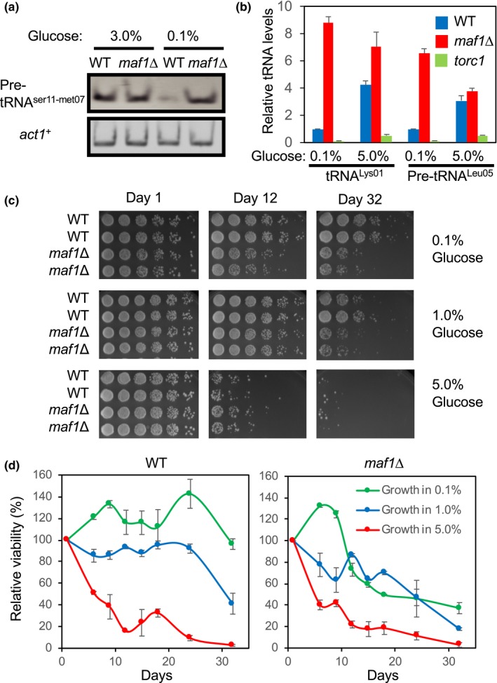
Figure 1.

Loss of Maf1 shortens chronological lifespan. (a) WT and maf1Δ cells were cultured in YES medium containing 3% or 0.1% glucose overnight at 30°C. Levels of the pre‐tRNAser11‐met07 and act1 + genes were examined by RT–PCR from total RNA preparation. tRNA samples were run on acrylamide gels and stained by SYBR Green. (b) WT and maf1Δ cells were cultured overnight in YES medium with 0.1% or 5.0% glucose. Expressions of the pre‐tRNALeu05 and tRNALys01 were examined by RT–PCR. The histograms show the relative tRNA expression levels normalized to act1+ expression. Data are expressed as the mean of three independent experiments. Error bars represent the standard error of the mean (SEM). (c) WT and maf1Δ cells were first cultured at 30°C in YES liquid medium with different percentages of glucose (0.1%, 1%, and 5%) for the indicated days. Fivefold serial dilutions of the cells were then plated on YES agar medium containing 3% glucose, incubated for 3 days at 30°C, and photographed. The complete presentation of the results is shown in Figure S2. (d) Quantification of the lifespan assays shown in Figure S2 was performed by using NIH Image J. The average growth intensity of each strain on each day was obtained from colonies derived from six dilutions. To calculate the average deviation (error bar), two strains for each genotype were tested, and their growth was quantified
