
Figure 6.
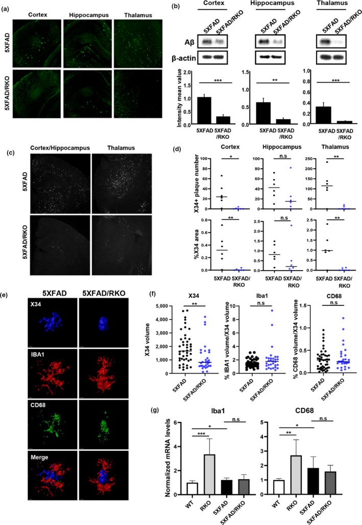
Deletion of REV‐ERBα mitigates amyloid plaque deposition in 5XFAD mice. (a) Representative image from thioflavin‐S staining of the brain sections including the cortex, hippocampus, and thalamus from 5XFAD and 5XFAD/REV‐ERα knockout (RKO) mice at 3.5 months. (n = 6–7 mice were analyzed per group). (b) Western blot analysis of Aβ peptide (4KDa) and β‐actin expression in each brain lysate. β‐actin was used as a loading control. **p < .01, ***p < .001 compared to the 5XFAD. (c) Representative image of X34 staining in the brain of 5XFAD and 5XFAD/RKO. (d) Quantification of X34‐positive plaque number and % area of X34 staining for each group mice brain using Image J. *p < .05, **p < .01 compared to the 5XFAD. (e) Representative images from confocal analysis of IBA1 and CD68 staining surrounding X34‐positive plaques in the cortex of 5XFAD and 5XFAD/RKO (X34 in Blue, IBA1 in Red, and CD68 in Green) (f) Quantification of X34‐positive plaque and plaque‐associated microglia (Iba1)/phagocytic microglia (CD68). Total volume of Iba1 and CD68 were normalized by X34 volume for each plaque. **p < .01 compared to the 5XFAD (n = 30–44 plaques) (g) mRNA expression of Iba1 and CD68 in the cortex of each group mice (WT, RKO, 5XFAD, 5XFAD/RKO). *p < .05, **p < .01, and ***p < .001