
Figure 7.
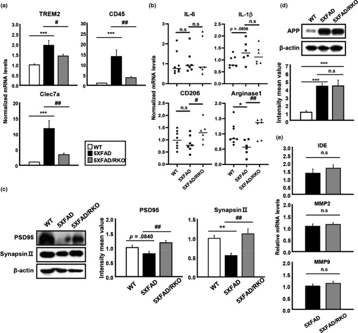
REV‐ERBα deletion in 5XFAD mice mitigates changes in DAM and synaptic markers and induces M2 microglial markers without alteration of APP processing. (a) mRNA expression of DAM markers including TREM2, CD45, and Clec7a and (b) pro‐inflammatory cytokines (IL‐6 and IL‐1β) as well as the M2 surface markers (CD206 and Arginase1) in the cortex of WT, 5XFAD, and 5XFAD/RKO. *p < .05, ***p < .001 compared to WT. # p < .05 and ## p < .01 compared to the 5XFAD. (c) Western blot analysis of synaptic markers PSD95 and synapsin II in the cortex of all three different genotypes of mice. β‐actin was used as a loading control. **p < .01 compared to WT and ## p < .01 compared to the 5XFAD. (d) Total amount of APP in the cortex of each group of mice by Western blot and (e) qPCR analysis of Aβ degradating enzymes (IDE, MMP2, and MMP9) from the same group of mice. ***p < .001 compared to WT