Figure 1.

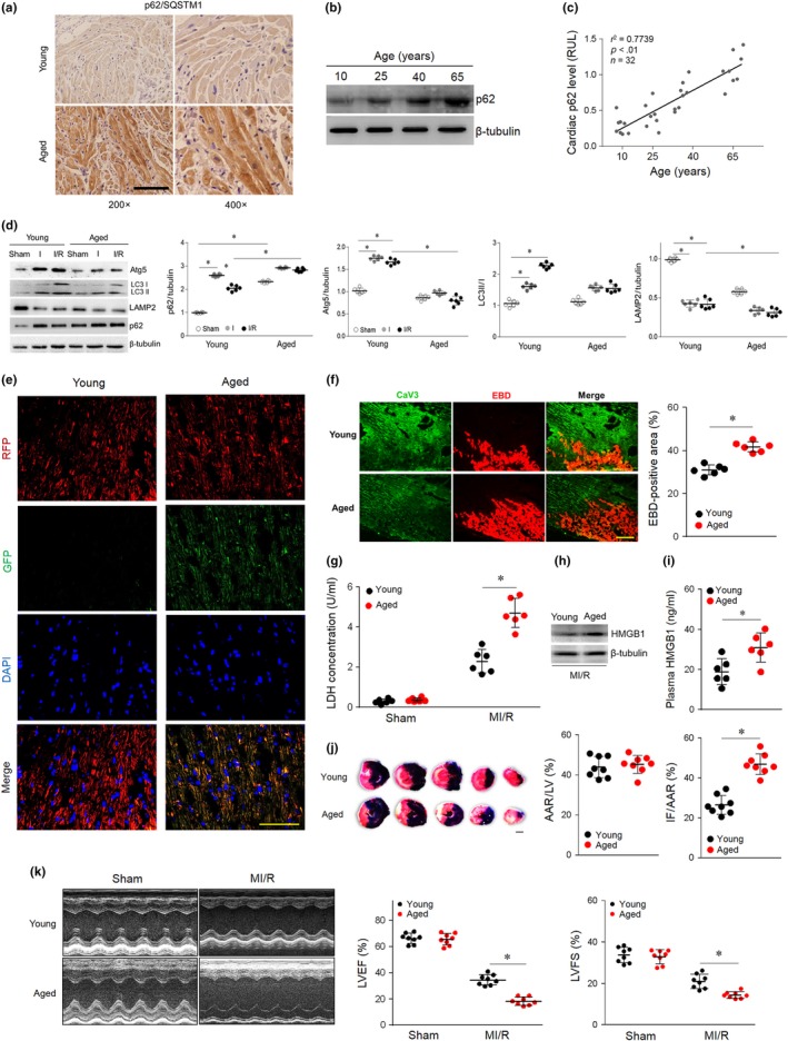
Impaired autophagic flux and enhanced necrosis. (a) Representative images of myocardial p62 immunohistochemical staining in young (10 years) and aged (65 years) human samples. (b) Representative Western blot for p62 protein levels in human myocardium samples from patients of different ages. (c) Linear regression analysis revealed a tendency for myocardial p62 to be positively related to age in the studied population. (d) Young and aged hearts were subjected to I/R (ischemia for 30 min and reperfusion for 120 min). Western blot analyses and quantification data of cardiac Atg5, LC3 II/I, LAMP2, and p62 levels. (e) Young and aged hearts were transfected with AAV9‐RFP‐GFP‐LC3 4 weeks prior to I/R surgery. Myocardial RFP‐GFP‐LC3 was stained and imaged by immunofluorescence. (f) Myocardial necroptosis was assessed by EBD uptake (red) in young and aged hearts subjected to ischemia (30 min) and reperfusion (240 min). (g) Serum LDH concentrations in each group. (h and i) Cardiac and plasma levels of HMGB1 in each group. (j) Area at risk (AAR) and infarct size (IF) in hearts subjected to I/R. (k) Average data for left ventricle ejection fraction (LVEF) and fractional shortening (LVFS) were assessed by echocardiography in young and aged mice subjected to sham or I/R injury (30 min of ischemia and 4 weeks of reperfusion). Scale bar = 20 μm. The values are the means ± SEM, n = 6 or 8 per group, *p < .05 versus the indicated groups
