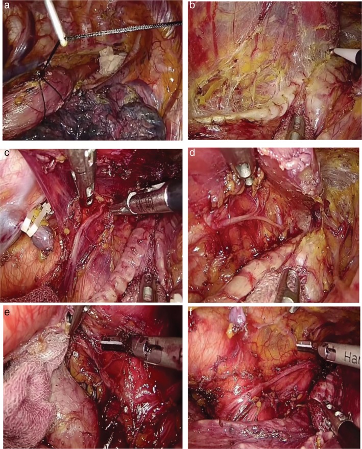
Figure 1.

Illustration of lymphadenectomy along the left RLN. (a) A crochet needle is punctured into the thorax to lift the esophagus with double 0# silk suture which has looped the esophagus at the level of the aortic arch. (b) The tracheoesophageal and primary esophageal arteries are identified in the esophageal mesenteriolum. (c) With good countertraction, the left RLN is clearly exposed and separated in the esophageal mesenteriolum above the level of the aortic arch. (d) The paratracheal lymph node is dissected and the left inferior thyroid artery is occasionally visible. (e) When the infra‐aortic arch lymph nodes have been dissected, the initial segment of the left RLN is confirmed. The trunk of the left pulmonary artery under the aortic arch is visible. (f) Following removal of the left RLN lymph nodes, the left RLN is easily clarified as running toward the oral side along the space between the lifted esophagus and trachea. A couple of superior cardiac branches of the sympathetic nerve system were identified. The thoracic duct is preserved.
