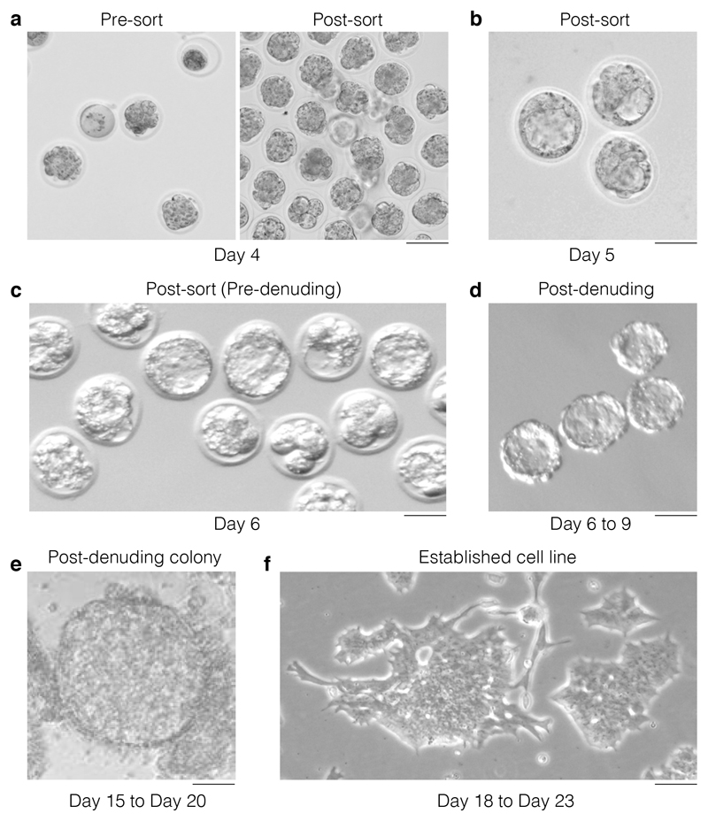
Figure 5. Morphological selection during the hESC derivation.
(a) On day 4 post harvesting and activation, embryos will be at a variety of developmental stages with some embryos looking sub-viable and some lysed (left panel: pre-sort). Upon removal of sub-viable or lysed embryos only the embryos that show viable characteristics are retained (right panel: post-sort). Scale bar 250 μm. (b) On day 5, the embryos that have developed to blastocyst and are suitable for zona pellucida removal are identified. Only select embryos that have a well expanded blastocoel for zona removal. Scale bar 250 μm. (c) The check and selection of blastocysts for zona removal is repeated on day 6 (representative images). Scale bar 250 μm. (d) Representative image depicting the morphologic aspect of blastocyst post-zona removal (days 6 to 9). These individual embryos are transferred to 96 well plates at this point. Scale bar 250 μm. (e) Representative image of a colony that has formed from a post-denuding blastocyst and that is ready for dissociation using trypsin. Scale bar 250 μm. (f) Representative image of an established hESC clone/cell line that is ready for quality control. Scale bar 250 μm.

