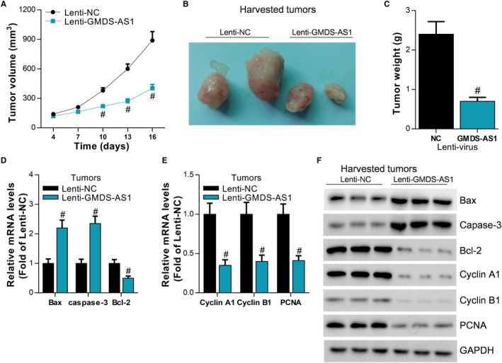
Figure 3.

GMDS‐AS1 inhibits LUAD cell proliferation in vivo. (A, B, C) PC‐9 cells stably overexpressing GMDS‐AS1 were injected subcutaneously into nude mice, and the growth of neoplasms was monitored in real time. After the nude mice were sacrificed, the neoplasms were removed and the weight was counted, # P < .05, compared with Lenti‐NC. (D, E) The expression levels of Bax, caspase‐3, Bcl‐2, cyclin A1, cyclin B1, and PCNA in neoplasms were detected by RT‐qPCR and Western blot, # P < .05, compared with Lenti‐NC
