
Figure 3.
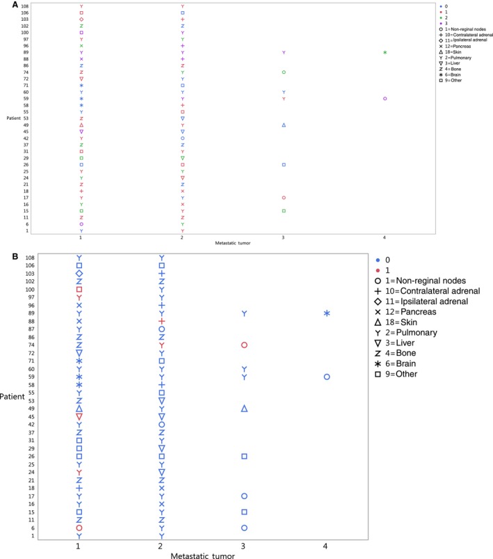
(A) PD‐1 IHC staining was performed on longitudinal metastatic tumors from 36 patients. PD‐1 staining is denoted as blue 0 = absent, red 1 = focal, green 2 = moderate, and purple 3 = marked. (B) PD‐L1 staining was performed on longitudinal metastatic tumors from 36 patients. Blue denotes PD‐L1 staining is absent and red denotes PD‐L1 staining is present. Symbols denote the metastatic location