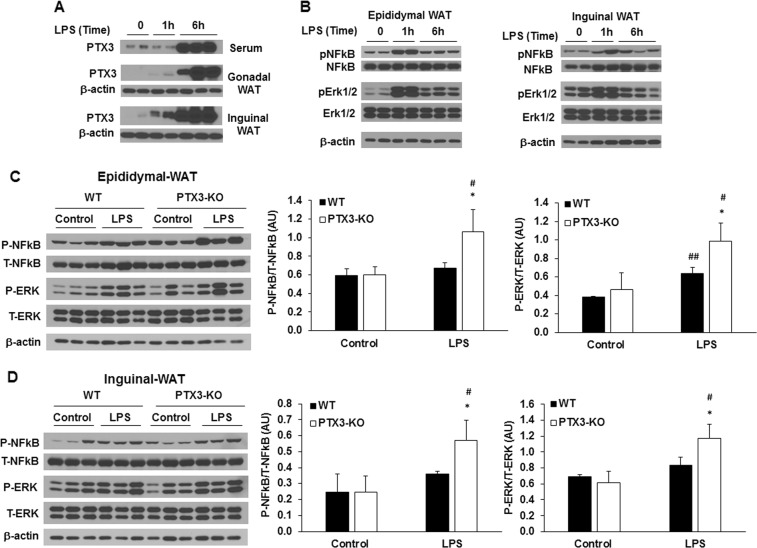
Fig. 6.
Effect of PTX3 deficiency on LPS-induced activation of inflammatory signaling pathways in adipose tissue. Time course of PTX3 protein expression (a) and activation of inflammatory signaling pathways (b) in adipose tissues treated with LPS in WT mice. Representative western blots for phosphorylated NF-κB p65 and phosphorylated Erk1/2 in epididymal (c) and inguinal (d) adipose tissue upon LPS treatment. n = 6 per group. The values are mean ± SD for the densitometric quantification of protein expression. *p < 0.05, **p < 0.01, ***p < 0.001 vs. WT; #p < 0.05, ##p < 0.01, ###p < 0.001 vs. control. WT: wild-type; KO: knockout

