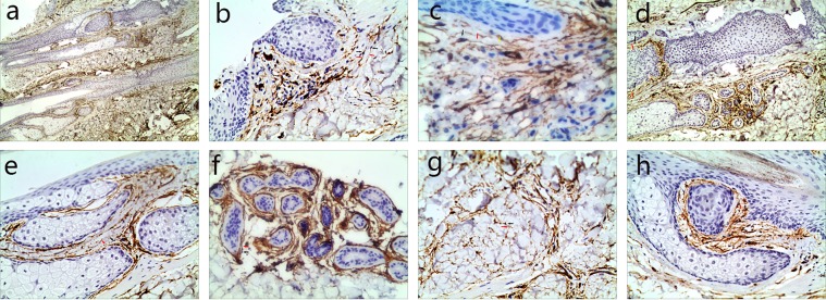
Figure 1.
Human scalp tissue stained immunohistochemically for CD34. (a) CD34+ cells (TCs) (black arrow) were in the deep dermis and subcutaneous tissue (40×). (b) A few TCs body (black arrows) with their Tps (red arrows) extended parallel to the adjacent epidermis (100×). (c) The spatial relationship of TCs body (black arrows) with their Tps (red arrows) and a blood vessel (yellow arrow) (100×). (d–g) TCs body (black arrows) and their Tps (red arrows) surrounded deep segments of HFs (d, 100×), sebaceous glands (e, 200×), secretory or ductal parts of the sweat glands (f, 200×) and intervals of adipose lobules (g, 200×). (h) TCs body (black arrows) and their Tps (red arrows) bordering the bulge and sub-bulge areas of HFs (200×).

