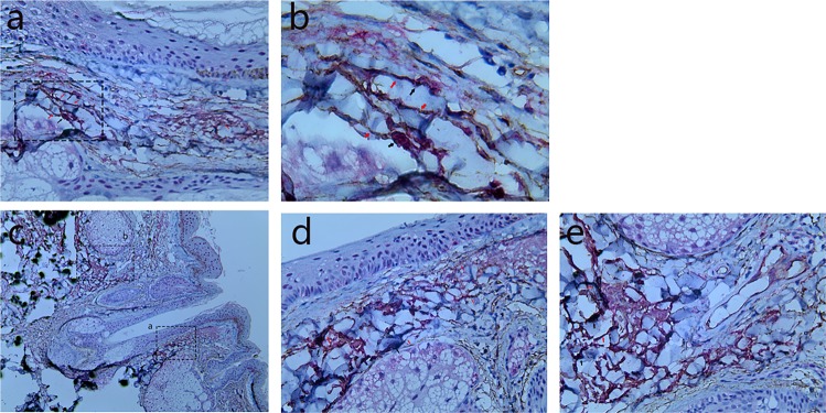
Figure 3.
Human scalp tissue stained immunohistochemically for CD117 and CD34. CD117+ cells (TCs body) (black arrows) with their Tps (red arrows) were observed in the reticular dermis, subcutaneous tissue of normal scalp (a, 200×; b, 400×), and around the middle of HFs (c, 100×; d, 200×; e, 200×). Figure (b) is an enlargement of the rectangular frame of Figure (a). Figure (d) is an enlargement of the rectangular frame a of Figure (c). Figure (e) is an enlargement of the rectangular frame b of Figure (c).

