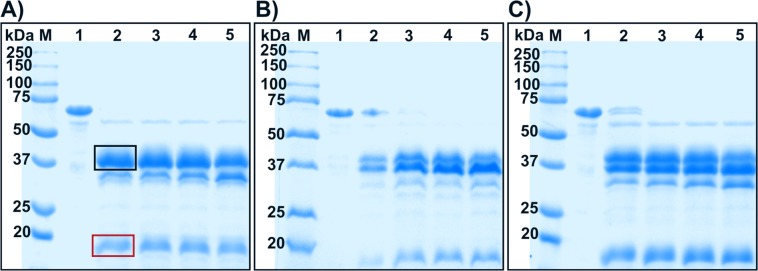
Figure 4.
SDS-PAGE gels of pro-jrPPO1-wt after digestion with A) trypsin, B) nagarse and C) proteinase K under reducing conditions. Lane M = marker, lane 1 = jrPPO1-wt without protease, lanes 2–5 = wild-type with protease (1% w/w) after 1, 2, 3 and 5 minutes. The red box highlights the still intact C-terminal domain and the black box highlights the band representing the active enzyme. Trypsin digestion (A) resulted in one sharp band for the active enzyme, thus indicating specific cleavage, whereas nagarse (B) and proteinase K (C) led to a more diffuse band pattern, indicating cutting at several positions. The gels were cropped to display the lanes of interest. (full-length gels: Fig. S24).

